External anatomy of a dogfish shark – The external anatomy of the dogfish shark, a fascinating species of cartilaginous fish, offers a glimpse into the remarkable adaptations that have shaped this ancient lineage. This comprehensive guide delves into the intricate details of the dogfish shark’s body, exploring its morphology, sensory structures, and unique characteristics.
From the streamlined body shape to the specialized fins and sensory organs, each aspect of the dogfish shark’s external anatomy plays a crucial role in its survival and success in the marine environment.
External Morphology of a Dogfish Shark

The dogfish shark, also known as the spiny dogfish, is a small species of shark commonly found in the northern Atlantic and Pacific Oceans. It has a distinctive appearance and several unique morphological features.
General Body Shape and Size
The dogfish shark has a slender, elongated body with a slightly flattened head and a pointed snout. Its body is covered in small, rough dermal denticles, giving it a sandpaper-like texture. Adult dogfish sharks typically range in size from 60 to 100 cm in length, with females generally being larger than males.
Fins
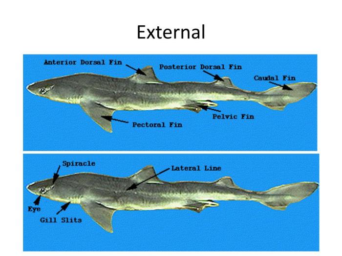
The dogfish shark possesses several fins that serve various functions. The dorsal fins are located on the back of the shark and help to stabilize and maneuver the body. The pectoral fins are located on the sides of the body behind the gills and are used for steering and balance.
The pelvic fins are located on the ventral side of the body behind the pectoral fins and are used for stability and maneuvering. The anal fin is located on the ventral side of the body behind the pelvic fins and helps to stabilize the shark while swimming.
Head and Sensory Structures: External Anatomy Of A Dogfish Shark

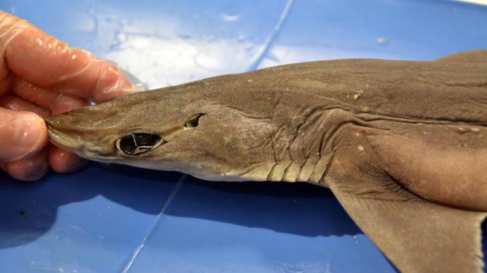
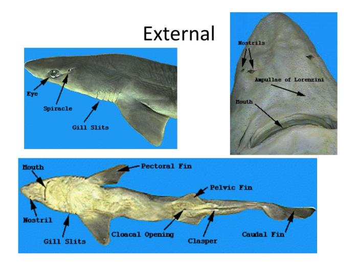
The dogfish shark’s head is dorsoventrally flattened and triangular in shape, with a pointed snout and large eyes located on the dorsal surface. The mouth is located ventrally, with a pair of nostrils positioned anteriorly. Spiracles, which are openings behind the eyes that lead to the respiratory system, are also present.
Eyes
The dogfish shark’s eyes are large and located on the dorsal surface of the head, providing binocular vision. They possess a tapetum lucidum, a reflective layer behind the retina, which enhances light sensitivity and aids in vision in dim conditions.
Nostrils, External anatomy of a dogfish shark
The nostrils are located anteriorly on the ventral surface of the head. They are used for olfaction, allowing the shark to detect chemical cues in the water.
Mouth
The mouth is located ventrally on the head. It is equipped with sharp, pointed teeth that are used for grasping and tearing prey. The teeth are arranged in multiple rows, with new teeth constantly replacing those that are lost.
Spiracles
Spiracles are openings located behind the eyes on the dorsal surface of the head. They lead to the respiratory system and allow water to enter the gills for respiration.
Sensory Organs
The dogfish shark possesses several sensory organs that aid in its survival. The lateral line system consists of a series of sensory cells located along the body that detect pressure changes in the water, allowing the shark to sense movement and vibrations.
Ampullae of Lorenzini are electroreceptors located around the head that can detect electrical fields generated by prey and other organisms.
Skin and Coloration
The skin of a dogfish shark is covered in small, tooth-like scales called placoid scales. These scales give the skin a rough texture and protect the shark from abrasion and injury.
Placoid scales are composed of a hard, bony material called dentin. The scales are arranged in a regular pattern, with each scale overlapping the one behind it. This arrangement creates a strong, flexible armor that protects the shark’s body.
Coloration
Dogfish sharks vary in color from brown to gray to black. The coloration of a dogfish shark is determined by a number of factors, including its diet, habitat, and age. Dogfish sharks that live in shallow water are often lighter in color than those that live in deep water.
This is because the lighter color helps to camouflage the shark from predators.
Dentition and Feeding Adaptations

The dogfish shark possesses a unique and specialized dentition that is adapted for capturing and consuming its prey. The teeth are arranged in rows on both the upper and lower jaws, with each row consisting of multiple cusps or points.
The teeth are sharp and pointed, enabling the shark to grasp and hold onto its prey securely.The jaws of the dogfish shark are also adapted for feeding. The upper jaw is protrusible, meaning it can be extended forward to increase the gape of the mouth.
This allows the shark to take in larger prey items. The hyoid arch, a structure located behind the jaws, also plays a role in feeding. It supports the tongue and helps to guide food into the esophagus.
User Queries
What is the function of the dorsal fin in a dogfish shark?
The dorsal fin provides stability and balance while swimming, preventing the shark from rolling over.
How do dogfish sharks use their ampullae of Lorenzini?
Ampullae of Lorenzini are electroreceptors that allow dogfish sharks to detect weak electrical fields emitted by prey, aiding in hunting.
What is the significance of placoid scales in dogfish sharks?
Placoid scales reduce drag and increase swimming efficiency, providing an advantage in the water.